
Identifying the Neuroinflammatory Transcriptomic Signature of Chemoradiotherapy-Induced Brain Injury Using GEO2R, KEGG, and GO Enrichment Analysis

ABSTRACT
Brain cancer damages the nervous system and is often treated with a combination of radiation therapy and chemotherapy. While effective against tumors, this treatment also damages healthy brain tissue, and most long-term survivors develop lasting problems with memory, attention, and planning, a condition with no approved treatment or prevention strategy. This decline is believed to be driven mainly by ongoing inflammation involving the brain’s immune cells, but the specific genes responsible are not clearly mapped out. This study analyzed a public gene expression dataset measuring 757 inflammation-related genes in mouse brain tissue, comparing untreated animals to animals treated with combined radiation and chemotherapy. Using an online differential expression tool, 108 genes were significantly altered between groups, and 27 were selected for pathway analysis based on statistical significance and magnitude of change. 3 genes were increased and 24 were decreased. The two most significantly changed individual genes, both markers of brain immune cell activation, were reduced in the treated group. Pathway analysis identified 65 significantly affected biological pathways, centered on immune receptor activity and a defense system called the complement system, alongside engulfment of cellular debris and cytokine production. The same core group of complement and immune receptor genes appeared repeatedly across these pathways, pointing to one coordinated response rather than scattered changes. These findings define a molecular signature of treatment-induced brain damage, offering a baseline for future studies testing whether stem cell-based therapies can reverse this damage and preserve cognitive function in brain cancer survivors.
INTRODUCTION
Brain cancer refers to malignant tumors of the brain and central nervous system, which cause a high mortality rate among all age groups due to their location and effect on neurological function [14]. It affects a substantial number of people each year, with a rising trend in the last decade [11].
Cranial radiotherapy, often combined with chemotherapy drugs like temozolomide (TMZ), is a standard brain cancer treatment [17]. While effective against tumors, it damages healthy brain tissue: 50–90% of long-term survivors develop lasting decline in memory, attention, and executive function, known as radiation-induced cognitive decline (RICD) [10]. In simple terms, curing the tumor can leave survivors with a permanently altered brain, which is a major quality-of-life problem in cancer care. No approved treatment currently prevents or reverses RICD [10]. The problem this research addresses is finding a safe way to protect the brain from chemoradiotherapy’s effects.
Evidence shows RICD is driven largely by sustained neuroinflammation. Radiation activates microglia (brain immune cells), triggers complement signaling, and raises inflammatory markers, damaging neuronal connections and impairing cognition [9]. Blocking this response preserves cognition in irradiated animals, confirming neuroinflammation as a treatable driver of RICD, not just a side effect [7].
Building on this, transplanting human neural stem cells (hNSCs) into irradiated brains reduces neuroinflammation and preserves cognition [1]. Because transplantation is invasive, researchers now use extracellular vesicles (EVs), particles secreted by hNSCs carrying protective cargo that reproduce much of this benefit without surgery [4, 16]. hNSC-EVs reduce microglial activation and restore cognition across several brain injury models [8, 16].
What remains poorly understood is the specific gene-level neuroinflammatory signature caused by chemoradiotherapy itself. It ends up being the baseline needed before EV-based rescue can be properly evaluated. This study used GEO2R differential expression analysis of a 757-gene neuroinflammation panel to identify genes significantly altered in mouse brain after chemoradiotherapy (RT+TMZ) versus untreated controls, characterizing the pathways involved via KEGG [13] and GO [5] enrichment. We hypothesize that chemoradiotherapy would significantly alter gene expression and key biological pathways in brain cancer tissue, and that these molecular changes can reveal biological pathways associated with treatment-related brain injury. This matters because defining that signature is a necessary first step toward understanding, and eventually counteracting, how chemoradiotherapy damages the brain. This research is important to the patients who deserve to live a long life without a trade-off on their mental capacity.
METHODS
Data Collection and Analysis of GEO2R Data
In this study, the dataset GSE319089, titled “Transcriptomic Effects of Human Neural Stem Cell Derived Extracellular Vesicle Supplementation Following Chemoradiotherapy for Brain Cancer” [6], was collected from the NCBI Gene Expression Omnibus (GEO) [3] using the GEO2R analysis tool. The dataset was identified using the keywords “brain cancer,” “chemotherapy,” and “homo sapien.” The dataset consists of expression profiling by array data from Mus musculus, measuring 757 genes related to neuroinflammation across male mouse brain tissue.
The dataset was then categorized into two groups: control (vehicle-treated, untreated) and chemoradiotherapy-treated (RT+TMZ, radiation combined with temozolomide), with 4 biological replicates in each group. These groups were defined and assigned within the no-code GEO2R bioinformatics tool, which uses the R programming language on the backend to perform differential expression analysis.
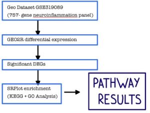
Figure 1: Research methodology: the steps and bioinformatics tools used in this study.
Identification of the Top Differentially Expressed Genes
To identify the most significant differentially expressed genes, statistical analysis was applied using GEO2R’s built-in limma-based linear modeling [15]. This process used adjusted p-value (adj. P < 0.05) in combination with log2 fold change to prioritize the most altered genes across samples in Google Sheets. A total of 108 genes met this significance threshold out of the 757 genes assayed on the neuroinflammation panel.
Functional and Enrichment Analysis Using SRPlot, KEGG, and GO Bioinformatics Tools
SRplot [18], KEGG [13], and GO [5] bioinformatics tools and databases were then utilized to analyze the functions of these top genes. These tools helped uncover the potential roles of the genes in complement activation and immune receptor activity pathways broadly associated with microglial activation and immune signaling in the chemoradiotherapy-treated brain.
RESULTS
Identification of Differentially Expressed Genes
GEO2R, the differential expression tool in NCBI’s Gene Expression Omnibus, was used to compare gene expression between control and chemoradiotherapy-treated (RT+TMZ) mouse brain samples across the 757-gene neuroinflammation panel. In the volcano plot (Figure 2), red dots represent significantly upregulated genes, blue dots represent significantly downregulated genes (both adj. P < 0.05), and black/grey dots represent genes that did not meet significance. From the accompanying Venn diagram, 115 of the 757 total genes on the panel were flagged as differentially expressed between the Control and ChemoRT groups, while the remaining 642 genes showed no significant difference.
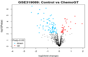
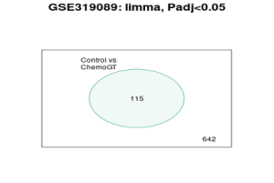
Figure 2: Volcano plot and Venn diagram of DEGs between Control and ChemoRT groups.
The two most significantly altered individual genes were Clec7a and Cd84, both markers of microglial activation, and both significantly downregulated in the chemoradiotherapy-treated group, indicating an early shift in innate immune gene expression following treatment.
Identification of the Top 27 Statistically Significant DEGs
Genes were narrowed to the most significant DEGs using adjusted p-value combined with log2 fold change, yielding 27 DEGs (3 upregulated, 24 downregulated) for pathway analysis. This gene list was submitted to SRplot for KEGG and Gene Ontology enrichment, using Mus musculus as the reference organism.
Potential Functions and Enrichment of the Identified Genes and Pathways
The KEGG analysis identified 65 significantly enriched pathways, the strongest of which centered on innate immune and phagocytic signaling, including complement and coagulation cascades, Toll-like receptor signaling, the phagosome pathway, efferocytosis, osteoclast differentiation, B cell receptor signaling pathway, and systemic lupus erythematosus. Genes encoding complement components (C3, C1qa, C1qb, C1qc) and Fc-receptors (Fcgr1, Fcgr2b, Fcgr3) recurred across nearly every top pathway, alongside Tlr2, Stat1, and Mapk12.
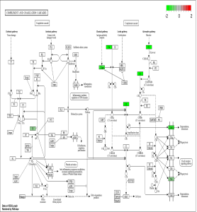
Figure 3: KEGG pathway diagram, Complement and Coagulation Cascades, with significantly altered genes shown in green (downregulated): C3, C1qrs, C4, C3AR1, CR4, PS.
Gene Ontology (GO) analysis. GO enrichment across all three ontologies reinforced this same immune and phagocytic signature. Enriched Biological Process terms included positive regulation of cytokine production, regulation of immune effector process, regulation of phagocytosis, regulation of leukocyte mediated immunity, and interleukin-1 beta production. Enriched Cellular Component terms included membrane raft, membrane microdomain, and phagocytic vesicle — locations directly tied to immune receptor clustering and signaling.
Enriched Molecular Function terms included immune receptor activity and immunoglobulin binding.
Figure 4: GO enrichment results across Biological Process, Cellular Component, and Molecular Function.
Conclusion
Together, these results show that chemoradiotherapy induces a predominantly suppressive shift in microglial, complement, and innate immune receptor gene expression in the mouse brain. Rather than a scattered set of unrelated changes, the same core genes, C3, C1qa/b/c, Tlr2, and the Fc-gamma receptors, appeared repeatedly across independent pathways, pointing to a coordinated neuroinflammatory effect. This supports the hypothesis that chemoradiotherapy would significantly alter gene expression and key biological pathways in brain cancer tissue, and that these molecular changes can reveal biological pathways associated with treatment-related brain injury. These findings establish a defined baseline against which the neuroprotective effects of hNSC-derived extracellular vesicle supplementation can now be meaningfully evaluated in future comparisons [6, 16].
DISCUSSION
Summary of Findings
The main goal of this study was to identify the gene-level neuroinflammatory signature produced by chemoradiotherapy in mouse brain tissue. From GEO2R analysis, the two most significantly altered genes were Clec7a and Cd84, both markers of microglial activation, and both significantly downregulated in the chemoradiotherapy-treated group. GO and KEGG enrichment identified 65 significantly enriched pathways total, many of which are shown in the pathway map (Figure 3).
Interpretation of Results
These results show a clear impact on complement and immune receptor signaling, with implications for cognitive function after chemoradiotherapy in Mus musculus. GO organizes genes by the biological role they play (grouped into Biological Process, Cellular Component, and Molecular Function), while KEGG maps genes onto specific, named biological pathways. This directly addresses the research question: radiation-induced cognitive decline (RICD) is understood to be driven by sustained neuroinflammation, and this study’s goal was to define the specific gene-level signature that chemoradiotherapy produces, so that signature can eventually be targeted.
Comparison with Previous Studies
Complement activation, the strongest and most recurring theme in this study’s KEGG results, has direct precedent as a driver of RICD specifically. One study [12] showed that glia-selective deletion of complement C1q prevents radiation-induced cognitive deficits and neuroinflammation in mice, directly demonstrating that the same complement genes altered in this dataset (C1qa/b/c) are not just associated with RICD but causally involved in producing it. More broadly, another study [20] describes how the C1q–C3–CR3 complement axis tags synapses for removal by microglia, providing the general mechanism by which the complement genes found here would translate into synaptic loss and cognitive impairment.
The immune receptor activity signature identified in this study (Fcgr1, Fcgr2b, Fcgr3, Tlr2) also has independent support in the literature. Ulvestad [19] established that Fc-gamma receptors on microglia mediate phagocytosis and cytotoxicity toward antibody-coated targets, meaning the Fcgr downregulation seen here plausibly reflects reduced microglial phagocytic capacity, not just reduced receptor expression. Babcock [2] showed that TLR2 is selectively upregulated by microglia in response to brain injury and drives the recruitment of immune cells to the site of injury, giving context for why Tlr2’s altered expression here is relevant to how the brain organizes its response to chemoradiotherapy-induced damage. Together, these four studies support treating this study’s complement and Fc/TLR receptor findings as parts of one interconnected innate immune response, consistent with how the genes clustered across pathways in the KEGG results.
Implications
Because complement C1q has already been shown to be a viable therapeutic target in RICD specifically [12], and because C5aR1 inhibition has independently been shown to alleviate cranial radiation-induced cognitive decline [7], this study’s finding that complement genes are centrally involved supports continued development of complement-targeted therapies (small-molecule inhibitors or antibody-based approaches against C1q, C3, or their receptors) as a strategy to prevent or reduce RICD in brain cancer patients.
Limitations
Since this study uses bioinformatics datasets from microarray experiments conducted by other researchers, one limitation is that the identified genes will need to be further studied in a laboratory or clinical environment before a treatment or prevention strategy can be developed for the brain damage that is a major trade-off of chemoradiotherapy.
Future Directions
The identified genes can be tested in the laboratory or in clinical trials to determine whether there is a way to stop the effects of chemoradiotherapy on the brain. Namely by restoring the complement and immune receptor signals and reducing cognitive impairment.
Acknowledgements
I would like to thank my family first for pushing me to go for things when they seem hard, because “nothing is impossible as long as you have an imagination.” Secondly, I’d like to thank Dr. Jedaidah Chilufya and Rising Researchers for allowing me the opportunity to be a part of such a noteworthy opportunity. Finally, I’d like to thank myself for continuing to try, even (especially) when it got hard.
References
- Acharya, M. M., Christie, L.-A., Lan, M. L., Giedzinski, E., Fike, J. R., Rosi, S., & Limoli, C. L. (2011). Human neural stem cell transplantation ameliorates radiation-induced cognitive dysfunction. Cancer Research, 71(14), 4834–4845.
- Babcock, A. A., Wirenfeldt, M., Holm, T., Nielsen, H. H., Dissing-Olesen, L., Toft-Hansen, H., Millward, J. M., Landmann, R., Rivest, S., Finsen, B., & Owens, T. (2006). Toll-like receptor 2 signaling in response to brain injury: An innate bridge to neuroinflammation. Journal of Neuroscience, 26(49), 12826–12837.
- Barrett, T., Wilhite, S. E., Ledoux, P., Evangelista, C., Kim, I. F., Tomashevsky, M., Marshall, K. A., Phillippy, K. H., Sherman, P. M., Holko, M., Yefanov, A., Lee, H., Zhang, N., Robertson, C. L., Serova, N., Davis, S., & Soboleva, A. (2013). NCBI GEO: Archive for functional genomics data sets—Update. Nucleic Acids Research, 41(D1), D991–D995.
- Baulch, J. E., Acharya, M. M., Allen, B. D., Ru, N., Chmielewski, N. N., Martirosian, V., Giedzinski, E., Syage, A., Park, A. L., Benke, S. N., Parihar, V. K., & Limoli, C. L. (2016). Cranial grafting of stem cell-derived microvesicles improves cognition and reduces neuropathology in the irradiated brain. Proceedings of the National Academy of Sciences, 113(17), 4836–4841.
- Gene Ontology Consortium. (2004). The Gene Ontology (GO) database and informatics resource. Nucleic Acids Research, 32(suppl_1), D258–D261.
- Hudson, C., Krattli, R. P., Jr., El-Khatib, S. M., Vagadia, A. R., et al. (2026). Human neural stem cell-derived extracellular vesicles improve cognitive function following glioma chemoradiation therapy. Cancer Letters.
- Krattli, R. P., Jr., Do, A. H., El-Khatib, S. M., Alikhani, L., Markarian, M., Vagadia, A. R., Usmani, M. T., Madan, S., Baulch, J. E., Clark, R. J., Woodruff, T. M., Tenner, A. J., & Acharya, M. M. (2026). C5aR1 inhibition alleviates cranial radiation-induced cognitive decline. Cancer Research, 86(1), 255–272.
- Leavitt, R. J., Acharya, M. M., Baulch, J. E., & Limoli, C. L. (2020). Extracellular vesicle-derived miR-124 resolves radiation-induced brain injury. Cancer Research, 80(19), 4266–4277.
- Lumniczky, K., Szatmári, T., & Sáfrány, G. (2017). Ionizing radiation-induced immune and inflammatory reactions in the brain. Frontiers in Immunology, 8, Article 517.
- Makale, M. T., McDonald, C. R., Hattangadi-Gluth, J. A., & Kesari, S. (2017). Mechanisms of radiotherapy-associated cognitive disability in patients with brain tumours. Nature Reviews Neurology, 13(1), 52–64.
- Manake, R., Phillips, V., Gangi, A., & Ravikumar, J. (2024). Trends in the incidence of brain cancer: An observational study. Cureus, 16(10), e72805.
- Markarian, M., Krattli, R. P., Jr., Baddour, J. D., Alikhani, L., Giedzinski, E., Usmani, M. T., Agrawal, A., Baulch, J. E., Tenner, A. J., & Acharya, M. M. (2021). Glia-selective deletion of complement C1q prevents radiation-induced cognitive deficits and neuroinflammation. Cancer Research, 81(7), 1732–1744.
- Ogata, H., Goto, S., Sato, K., Fujibuchi, W., Bono, H., & Kanehisa, M. (1999). KEGG: Kyoto Encyclopedia of Genes and Genomes. Nucleic Acids Research, 27(1), 29–34.
- Ostrom, Q. T., Francis, S. S., & Barnholtz-Sloan, J. S. (2021). Epidemiology of brain and other CNS tumors. Current Neurology and Neuroscience Reports, 21(12), 68.
- Ritchie, M. E., Phipson, B., Wu, D., Hu, Y., Law, C. W., Shi, W., & Smyth, G. K. (2015). Limma powers differential expression analyses for RNA-sequencing and microarray studies. Nucleic Acids Research, 43(7), e47.
- Smith, S. M., Giedzinski, E., Angulo, M. C., Lui, T., Lu, C., Park, A. L., Tang, S., Martirosian, V., Ru, N., Chmielewski, N. N., Liang, Y., Baulch, J. E., Acharya, M. M., & Limoli, C. L. (2020). Functional equivalence of stem cell and stem cell-derived extracellular vesicle transplantation to repair the irradiated brain. Stem Cells Translational Medicine, 9(1), 93–105.
- Stupp, R., Mason, W. P., van den Bent, M. J., Weller, M., Fisher, B., Taphoorn, M. J. B., Belanger, K., Brandes, A. A., Marosi, C., Bogdahn, U., Curschmann, J., Janzer, R. C., Ludwin, S. K., Gorlia, T., Allgeier, A., Lacombe, D., Cairncross, J. G., Eisenhauer, E., & Mirimanoff, R. O. (2005). Radiotherapy plus concomitant and adjuvant temozolomide for glioblastoma. New England Journal of Medicine, 352(10), 987–996.
- Tang, D., Chen, M., Huang, X., Zhang, G., Zeng, L., Zhang, G., Wu, S., & Wang, Y. (2023). SRplot: A free online platform for data visualization and graphing. PLOS ONE, 18(11), e0294236.
- Ulvestad, E., Williams, K., Matre, R., Nyland, H., Olivier, A., & Antel, J. (1994). Fc receptors for IgG on cultured human microglia mediate cytotoxicity and phagocytosis of antibody-coated targets. Journal of Neuropathology & Experimental Neurology, 53(1), 27–36.
- Zong, S., Cui, X., Wu, S., … Lu, Z. (2026). Microglia and neuroinflammation: Function, heterogeneity, and crosstalk. Cellular & Molecular Immunology, 23(7), 787–805.


